Science
University of Victoria Breakthrough Revolutionizes Microscopy
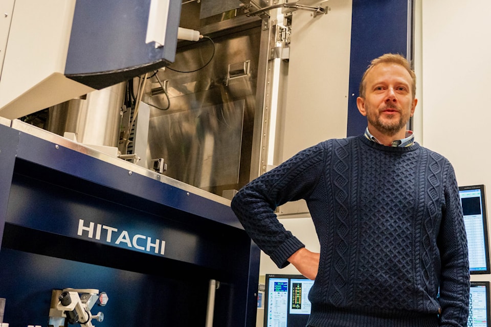
A team of researchers at the University of Victoria has made a significant advancement in the field of electron microscopy. This breakthrough enables scientists to visualize atomic-scale structures using more accessible, lower-cost equipment. Under the leadership of Arthur Blackburn, co-director of the university’s Advanced Microscopy Facility, the team developed a novel imaging technique that achieves sub-Ångström resolution—less than one ten-billionth of a metre.
Traditionally, such high-resolution imaging required large and expensive transmission electron microscopes (TEM). However, this new method employs a compact, low-energy scanning electron microscope (SEM) paired with advanced computational techniques. “This work shows that high-resolution imaging doesn’t have to rely on expensive, complex equipment,” Blackburn stated. As the Hitachi High-Tech Canada Research Chair in Advanced Electron Microscopy, he emphasized the potential of this innovation to democratize access to cutting-edge microscopy.
The research, published in Nature Communications, highlights how this technique opens doors for laboratories worldwide. The approach allows for high-resolution imaging without the prohibitive costs, space requirements, and specialized personnel typically associated with traditional methods. By utilizing overlapping patterns of scattered electrons, the team constructed highly detailed images of samples, achieving a remarkable resolution of just 0.67 Ångström. This size is even smaller than that of an atom and is 1/10,000 the width of a human hair.
This advancement is poised to have transformative effects across various fields, particularly in materials science, nanotechnology, and structural biology. Blackburn noted, “The advance will most immediately benefit the research and production of 2D materials, which are promising in the development of next-generation electronics.” Furthermore, the technique could assist in determining the structures of small proteins, potentially leading to breakthroughs in health and disease research.
Overall, the development at the University of Victoria stands as a pivotal moment in microscopy, offering unprecedented opportunities for scientific exploration and innovation. With this achievement, researchers can now explore atomic structures with greater ease and efficiency, fundamentally shifting the landscape of microscopic imaging.


Urgent Call for $150M Federal Funding for Qikiqtarjuaq Port

Saskatchewan Government Adds $1 Billion to Deficit – Urgent Update

Thomson and Weston Families Secure Hudson’s Bay Charter for $18M

Family of Colombian Man Challenges U.S. Military Strikes After Death

Bloc Québécois Aims to Amend Bill C-9, Targeting Hate Speech Exemptions

Family and Friends Mourn the Loss of Donald James Wonnacott

Hollywood Stars Dine at South Osborne’s Vera Pizzeria

Calgary Teen Charged for Allegedly Creating AI-Generated Explicit Images

Dental Oversight Puts Niagara Detention Centre Inmates at Risk

Secwepemc First Nation Seeks Aboriginal Title Over Kamloops Area

Scientists Unearth Ancient Antarctic Ice to Unlock Climate Secrets

Trump and McCormick to Announce $70 Billion Energy Investments

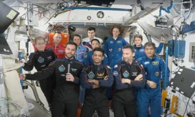
Four Astronauts Return to Earth After International Space Station Mission
TransLink Launches Food Truck Program to Boost Revenue in Vancouver

Apple Notes Enhances Functionality with Markdown Support in macOS 26

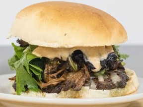
Manitoba’s Burger Champion Shines Again Amid Dining Innovations

Urgent Update: Fatal Crash on Highway 99 Claims Life of Pitt Meadows Man

Ukrainian Tennis Star Elina Svitolina Faces Death Threats Online
-
 Politics4 weeks ago
Secwepemc First Nation Seeks Aboriginal Title Over Kamloops Area
-
 World5 months ago
Scientists Unearth Ancient Antarctic Ice to Unlock Climate Secrets
-
 Entertainment5 months ago
Trump and McCormick to Announce $70 Billion Energy Investments
-
 Science5 months ago
Four Astronauts Return to Earth After International Space Station Mission
-

 Lifestyle5 months ago
TransLink Launches Food Truck Program to Boost Revenue in Vancouver
-
 Technology3 months ago
Apple Notes Enhances Functionality with Markdown Support in macOS 26
-
 Lifestyle3 months ago
Manitoba’s Burger Champion Shines Again Amid Dining Innovations
-
 Top Stories2 months ago
Urgent Update: Fatal Crash on Highway 99 Claims Life of Pitt Meadows Man
-
 Politics4 months ago
Ukrainian Tennis Star Elina Svitolina Faces Death Threats Online
-
 Sports5 months ago
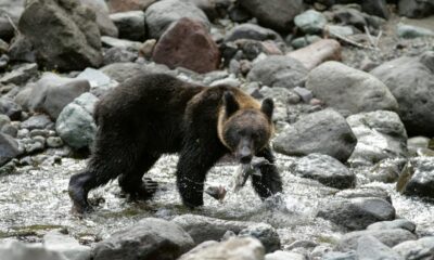
Search Underway for Missing Hunter Amid Hokkaido Bear Emergency
-

 Politics5 months ago
Carney Engages First Nations Leaders at Development Law Summit
-

 Technology5 months ago
Frosthaven Launches Early Access on July 31, 2025